
A tooth that
never appeared
can still cause trouble.
A dentigerous cyst can develop around an unerupted tooth inside the jaw. It may quietly expand and weaken nearby bone before there is an obvious lump or change in your pet’s behavior.
! If a tooth seems missing, especially in a young dog, ask your veterinarian whether dental imaging is needed to find out where it is.
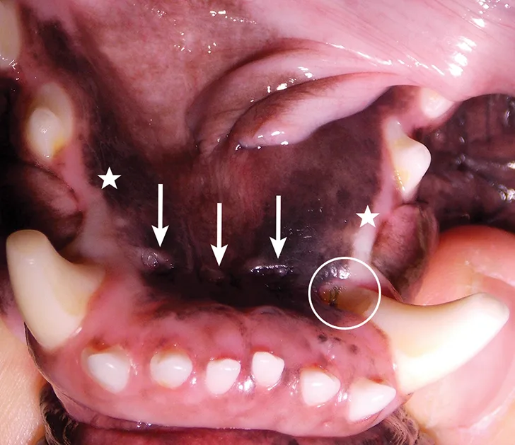
hidden risk
Sometimes the clue
is a tooth that’s missing.
Many dentigerous cysts are discovered on dental radiographs before a pet shows a visible problem. As a cyst grows, it may expand the jaw or shift nearby teeth.
A missing tooth does not automatically mean a cyst. The tooth may be absent, unerupted, impacted, or difficult to identify without imaging.
Arrange a veterinary exam for new oral swelling. Rapid enlargement, bleeding, marked pain, trouble eating, or breathing difficulty needs prompt veterinary attention.
The cyst develops around the crown of an unerupted tooth and may expand into the jawbone.
A fluid-filled sac
around a hidden tooth.
“Dentigerous” means associated with a tooth. The cyst forms around the crown of a tooth that has not erupted. It is not the same as an abscess, and other jaw lesions can look similar—so the diagnosis needs veterinary assessment.
It may be embedded in the jaw or blocked from erupting. The mouth can look almost normal.
Fluid accumulates in the tissue surrounding the unerupted tooth.
As it expands, the lesion can displace adjacent teeth and weaken the jawbone.
“It never came in”
is worth checking.
In dogs and cats, a tooth that is not visible may be absent—or it may still be inside the jaw. Dental radiographs can locate unerupted teeth and look for changes around them.
Not every unerupted tooth has a cyst. But identifying it gives the veterinary team a chance to discuss treatment or an appropriate monitoring plan before a hidden problem grows.
A quick look can count the teeth you see. Imaging can find the one you don’t.
Tell your veterinarian if a tooth seems missing, especially if there is swelling, a change in the bite, or a lump along the jaw.
Find the tooth.
Understand the lesion.
Dental radiographs show unerupted teeth and changes in the surrounding bone. Depending on the size and location, the veterinarian may recommend additional imaging, sampling, or histopathology to confirm what the lesion is.
Veterinarian? Discuss a caseA diagnostic workup may include
Oral exam and tooth chart
Document visible teeth, swelling, bite changes, and any missing
tooth positions.
Intraoral dental radiographs
Locate the unerupted tooth and assess the surrounding jawbone and
adjacent roots.
Advanced imaging when needed
CT may help define a large or complex lesion and support surgical
planning.
Laboratory confirmation
Tissue may be submitted for histopathology because radiographic
appearance alone cannot identify every lesion with certainty.
Remove the source.
Help the jaw heal.
Treatment depends on the cyst’s size, location, and effect on the jaw and nearby teeth. Surgery commonly removes the unerupted tooth and cyst lining; larger lesions may need a staged plan.
Map the affected area
Imaging helps the team understand the lesion’s boundaries and its relationship to roots, nerves, and surrounding bone.
Remove the tooth and cyst tissue
The unerupted tooth and cyst lining are typically removed surgically. A tissue sample may be sent to a veterinary pathologist.
Check healing over time
Rechecks and follow-up imaging may be recommended to assess bone healing and confirm the treatment site is recovering as expected.
Some larger cysts need staged treatment. The care team may first decompress a lesion to reduce its size before definitive removal. The approach is individualized to the pet and the anatomy involved.
Not seeing a tooth?
Let’s find out where it is.
If your veterinarian has found a missing or unerupted tooth, jaw swelling, or a suspected cyst, DVDP can help explain the dental evaluation and coordinate with your primary veterinary team.
Delaware Veterinary Dental Practice · Wilmington and Dover, DE · (302) 463-2879
Questions pet parents ask.
What causes a dentigerous cyst?
A dentigerous cyst forms around the crown of an unerupted tooth. Not every unerupted tooth develops a cyst, but a veterinarian may recommend imaging to locate it and assess the surrounding bone.
Can my pet have a dentigerous cyst without a visible lump?
Yes. Some are found incidentally on dental radiographs before an obvious swelling appears. Other pets may develop an expanding area along the jaw or gums.
Does a missing tooth mean my pet has a cyst?
No. The tooth may be absent or unerupted, and a cyst is only one possible concern. Dental radiographs help locate the tooth and evaluate the area.
Can a dental X-ray confirm a dentigerous cyst?
Radiographs can show an unerupted tooth and a surrounding lesion, but other conditions may look similar. The veterinarian may recommend tissue sampling and histopathology for confirmation.
How are dentigerous cysts treated?
Treatment commonly involves surgical removal of the unerupted tooth and cyst lining. Larger or more complex lesions may require additional imaging or a staged surgical approach.
Is a dentigerous cyst urgent?
Arrange veterinary assessment for a missing tooth with swelling or a suspected jaw lesion. Seek prompt care for rapidly enlarging swelling, bleeding, marked pain, trouble eating, or breathing difficulty.