The hidden risk behind a broken tooth
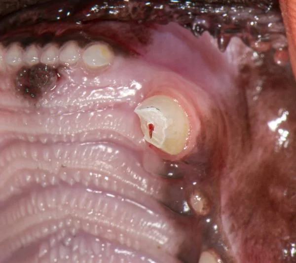
That little red dot could change everything.
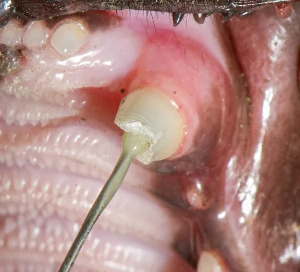
A broken tooth may look like a small chip. But if the living center is exposed, bacteria can reach deep inside the tooth. Your pet may keep playing, eating, and wagging while a painful problem develops. Prompt assessment can matter.
The part you can’t see
A tooth is alive on the inside.
At the center of every tooth is the pulp: soft tissue containing nerves and blood vessels. A fracture that opens a path to this living core gives oral bacteria a way in.
The result can be inflammation, loss of the tooth’s vitality, and infection around the root. The outside break may be only the first clue.
See the treatment paths
What to look for
Six clues that deserve a closer look.
You may see a clear fracture. Or you may only notice a small change in color, chewing, or mood. Any one of these signs is a reason to ask your veterinarian about the tooth.
A chipped or broken tooth
A jagged edge, shortened crown, or missing piece of tooth.
A red, pink, or dark spot
A dot or opening in the center of the broken surface may be pulp.
A tooth changing color
Pink, purple, gray, green, or dull discoloration can follow injury.
Different chewing habits
Dropping food, chewing on one side, or avoiding hard food and toys.
Face or mouth sensitivity
Pawing, drooling, flinching, bad breath, or resisting touch near the mouth.
No obvious behavior change
Pets can hide mouth pain. Eating normally does not clear the tooth.
If your pet is comfortable, take a photo to show your veterinarian. Don’t force the mouth open or touch the broken area.
The anatomy, made simple
The red center is small. The consequences can spread.
Think of the tooth as a protected structure. Enamel and dentin shield the pulp. When a fracture breaks through those layers, bacteria have a route toward the root.
- Enamel: the hard outer crown covering.
- Dentin: the layer beneath enamel surrounding the pulp.
- Pulp: living tissue containing nerves and blood vessels.
- Root: the part anchored beneath the gumline.
Why timing matters
A fracture doesn’t stay a harmless little chip.
When pulp is exposed, the tooth’s inner tissue is vulnerable. A veterinary exam can identify the damage and determine what treatment is appropriate.
It may be considered for selected recent fractures, often within 24–48 hours.
Root canal therapy or extraction may still be appropriate, depending on the tooth.
The 24–48-hour timeframe is commonly cited for considering vital pulp therapy. It is not a universal deadline for all treatment.
Treatment is not one-size-fits-all
There are paths forward. The tooth decides which one.
An anesthetized oral exam and dental radiographs help the veterinary team assess the fracture, root, and surrounding tissues.
Vital pulp therapy
For selected recent injuries, the affected portion of the pulp is removed and the remaining living tissue is protected and sealed. Follow-up imaging matters.
Root canal therapy
The pulp is removed, the canals are cleaned and sealed, and the opening is restored. This can preserve the tooth’s shape and function.
Surgical extraction
When the fracture or disease makes saving the tooth unsuitable, surgical removal treats the affected tooth and allows the site to heal.
Questions pet parents ask
Before you close this page, know this.
My pet seems fine. Could a broken tooth still be painful?
Yes. Pets can hide signs of mouth pain and continue eating. Normal behavior does not rule out a painful or damaged tooth.
Does every fractured tooth need to be extracted?
No. Depending on the fracture and the tooth’s condition, the veterinarian may consider vital pulp therapy, root canal therapy, or extraction. An examination and dental radiographs help guide the decision.
Is 48 hours a hard deadline?
No. The 24–48-hour period is commonly cited when considering vital pulp therapy for selected recent injuries. Root canal therapy or extraction may still be options after more time has passed.
What should I do while arranging an appointment?
Call your veterinarian for guidance. Avoid hard chews, don’t probe or try to seal the tooth, and don’t give human pain medication. Ask a veterinarian what is safe for your pet.
Delaware Veterinary Dental Practice
Found a broken tooth? Let’s get a clearer picture.
Contact your veterinarian promptly and ask whether a veterinary dental assessment is recommended. A small visible mark can hide a bigger problem below the surface.
Veterinarians: talk through a challenging case
Contact DVDP to discuss a dental case with our board-certified veterinary dentist and anesthesiologist.
This page is educational and cannot diagnose your pet. A veterinarian must examine the mouth and obtain appropriate dental imaging to recommend care.